tree in bud opacities pneumonia
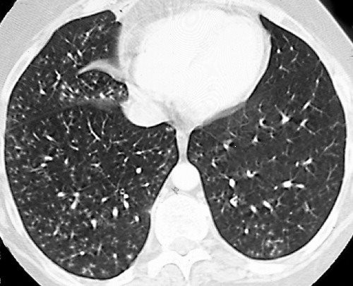
An example of the TIB pattern in a patient with COPD. This pattern is often seen in patients with chronic obstructive pulmonary disease COPD.
View Of Tree In Bud The Southwest Respiratory And Critical Care Chronicles
This may prompt inappropriate treatment.

. Tree-in-bud TIB opacities are a common imaging finding on thoracic CT scan. The patient underwent CT scanning of the chest which showed areas of nodular infiltration in the lower lobes with tree. Differential diagnosis of the tree-in-bud pattern includes com-munity-acquired as acute disease and mycobacterial pneumonia diffuse panbronchiolitis and DAB as subacute or chronic disease.
The patients chest radiograph is shown in the upper left corner. Thus pulmonary infiltrates can be mistaken as pneumonia as patients are considered immunocompromised. We tentatively suspected mycobacterial pneumonia based on tree-in-bud opacities on HRCT and Mycobacterium acid-fast staining Gaffky score 1.
CT finding of centrilobular nodules with TIB opacities was first described in pulmonary tuberculosis and is considered highly predictive of. Patients with aspiration pneumonia are some-times complicated with Mycobacterium infections especially elderly patients. Since the initial report of endobronchial spread of pulmonary tuberculosis the tree-in-bud sign has been reported in a wide variety of health conditions including infectious diseases aspiration pneumonia congenital disorders idiopathic disorders inhalation immunologic disorders connective disorders 23456 and central lung cancer involving the.
Histology of the open lung biopsy shows an organising pneumonia with extensive endobronchiolar granulation tissue arrows. HR-CT scan shows multiple centrilobular nodules and branching linear opacities causing the tree in bud pattern next to a pneumomediastinum. TIB opacities typically show branching configurations from secondary pulmonary lobules with sparing of subpleural lungs on CT thorax.
The appearance of a tree in bud is depicted by a pattern of bronchial dilatation and filling on a thin-section chestCT. The differential for this finding includes malignant and inflammatory etiologies either infectious or sterile. Usually somewhat nodular in appearance the tree-in-bud pattern is generally most pronounced in the lung periphery and associated with abnormalities of the larger airways.
More extensive lympho - cytic infiltrations may be associated with lymphoid interstitial pneumonia LIP with ground-. 1 2 3 4 Reported causes include infections aspiration and a variety of inflammatory conditions. In radiology the tree-in-bud sign is a finding on a CT scan that indicates some degree of airway obstruction.
1 The tree-in-bud sign is a nonspecific imaging finding that implies impaction within bronchioles the smallest airway passages in the lung. Tree-in-bud TIB opacities are a subset of centrilobular nodules. These small clustered branching and nodular opacities represent terminal airway mucous impaction with adjacent peribronchiolar inflammation.
A chest radiograph showed bilateral nodular opacities with a left lower lobar consolidative opacity Fig 1A 1B. Can pneumonia cause tree-in-bud. Tree-In-Bud Pattern A lymphoid interstitial infiltrate in the walls of the small airways follicular bronchiolitis may cause small centrilobular nodules and the tree-in-bud pattern Fig.
87 rows In the acute phase bacterial pneumonia manifests in the form of segmental or lobar. To the best of our knowledge the tree-in-bud sign on CT scan with biopsy proven bronchiolitis from cellular infiltration has not been described before. Tree-in-bud refers to a pattern seen on thin-section chest CT in which centrilobular bronchial dilatation and filling by mucus pus or fluid resembles a budding tree.
Nodular opacities with tree-in-bud appearance can be associated with other changes in lung parenchyma-such as thickening of the bronchial walls consolidations andor areas of.
2

Pdf Tree In Bud Semantic Scholar

Pdf Tree In Bud

Tree In Bud Pattern Pulmonary Tb Eurorad

Tree In Bud Pattern Radiology Case Radiopaedia Org
2
2

Tree In Bud Pattern Radiology Case Radiopaedia Org
2
Chest Ct With Multifocal Tree In Bud Opacities Diffuse Bronchiectasis Download Scientific Diagram

Co Rads 2 With Tree In Bud Sign A 27 Year Old Male Attended The Download Scientific Diagram

Tree In Bud Sign And Bronchiectasis Radiology Case Radiopaedia Org

Tree In Bud Pattern Pulmonary Tb Eurorad
View Of Tree In Bud The Southwest Respiratory And Critical Care Chronicles
Tree In Bud Sign Lungs
2

Hrct Scan Of The Chest Showing Diffuse Micronodules And Tree In Bud Download Scientific Diagram
2

Tree In Bud Pattern Pulmonary Tb Eurorad